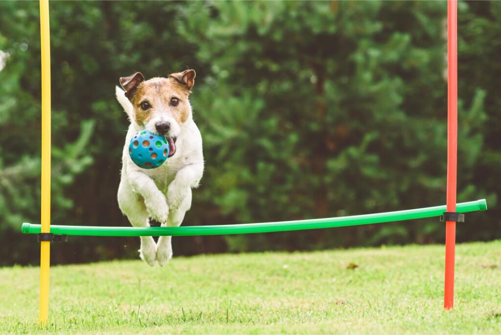
A “trick knee” in dogs—formally called a luxating patella—happens when the kneecap slides out of its groove. It’s common in small breeds, increasingly recognized in athletic and large dogs, and ranges from a mild annoyance to a life-limiting orthopedic problem. The good news: with the right plan—weight management, activity adjustments, targeted exercises, supplements, and, when needed, surgery—most dogs return to comfortable, confident movement.
This comprehensive guide explains luxating patella grading (I–IV), early signs, how vets diagnose it, non-surgical vs. surgical options, at-home rehab, braces and home modifications, and evidence-informed supplements.
One of the reasons I started this website is because I know what it’s like to leave the veterinary clinic with more questions than answers. Even with animal science degrees and a career in veterinary medicine, I’ve found myself going home after appointments and spending hours reading veterinary journals, talking with specialists, and trying to learn everything I could to help my own pets.
Over the years, I’ve cared for animals with complex medical conditions, and I understand how overwhelming it can feel when you’re trying to make the best decisions for a pet you love. That’s why I write these guides—to combine my education, experience, and personal research into one place that’s easy for pet owners to understand.
This article isn’t a substitute for your veterinarian’s advice, but my goal is to help you better understand the condition, know what questions to ask, and learn about the treatments, diets, supplements, and products that may help improve your dog’s quality of life.
What Is a Luxating Patella?
The patella (kneecap) normally tracks in a groove on the femur. With a luxating patella (medial is most common), the kneecap pops in and out of that groove—usually inward (medial), sometimes outward (lateral). Each slip irritates cartilage, destabilizes the knee, and can contribute to pain, arthritis, and secondary problems like cranial cruciate ligament (CCL) tears.
Breeds often affected:
- Small breeds: Pomeranian, Chihuahua, Yorkshire Terrier, Maltese, Pug, French Bulldog, Boston Terrier, Mini Poodle
- Also seen in active/large breeds and mixes, especially with straight or shallow stifles
Contributing factors include heredity, limb alignment, shallow trochlear grooves, tight/imbalanced soft tissues, and trauma.
Patellar Luxation Grades (I–IV)
Veterinarians grade severity during exam. Knowing the grade helps guide treatment.
- Grade I: Kneecap sits in the groove but can be manually pushed out; pops back on its own. Often minimal signs.
- Grade II: Kneecap pops out on its own at times and can be put back; intermittent lameness or “skipping.”
- Grade III: Kneecap is out most of the time but can be pushed in; moderate, consistent lameness and deformity develops.
- Grade IV: Kneecap is permanently out and cannot be reduced; severe deformity/arthritis and significant lameness.
Grades I–II are often managed conservatively; Grades III–IV frequently need surgery—especially in young or symptomatic dogs.
Early Signs and Symptoms
Watch for:
- “Skipping” gait—your dog briefly holds up a back leg, then resumes normal stride
- Sudden yelp or hop during play, then normal a moment later
- Bow-legged stance (medial luxation) or knock-kneed changes (lateral)
- Difficulty jumping, reluctance to use stairs, or hesitation to rise
- Sitting with one leg kicked out to the side
- Thigh muscle loss over time; worsening after activity
Catching symptoms early can slow long-term joint damage.
How Vets Diagnose Luxating Patella
- Orthopedic exam: Palpation reveals patellar tracking and how easily it luxates; assessment for pain, effusion, crepitus.
- Radiographs (X-rays): Evaluate bone alignment, arthritis; rule out fractures.
- Alignment and limb conformation: Tibial torsion, femoral varus/valgus, shallow trochlear groove.
- Additional imaging (advanced cases): CT or 3D planning when significant bone realignment is considered.
If your dog also shows instability or drawer sign, your vet will consider concurrent CCL disease, which changes the plan.
Non-Surgical Management (Often First-Line for Grades I–II)
Many dogs—especially mild cases—do well with a focused conservative plan. Key goals: improve patellar tracking, strengthen the “quads core,” reduce inflammation, and protect cartilage.
1) Keep Your Dog Lean (the single most powerful step)
- Target a body condition score (BCS) 4–5/9. Even a 5–10% weight loss lightens joint load and reduces luxation frequency.
- Feed measured meals; use high-protein, controlled-calorie diets; swap high-calorie treats for training kibble.
High-protein weight-management dog food
2) Activity Optimization
- Do: Daily, low-impact, consistent movement—leashed walks on grass, controlled hills, gentle strengthening drills.
- Avoid: Sudden sprints, repetitive jumping on/off furniture, slippery floors, frantic fetch with rapid stops/turns.
- Use a padded, Y-front harness to protect the neck and encourage balanced gait.
3) Home Modifications
- Non-slip runners over hard floors (kitchen, hallways).
- Stairs/sofa/car ramps; block bed/couch jumping.
- Orthopedic bed with supportive foam and side bolsters for alignment.
4) Physical Rehabilitation (Canine PT)
A rehab therapist can build a safe, progressive strengthening program to stabilize the patella and improve limb alignment.
Useful exercises (introduce with professional guidance):
- Sit-to-stand (slow, square sits): 5–10 reps, 1–2 sets daily
- Controlled step-ups on a low, stable platform: 5–10 reps each hind leg
- Weight shifting: Stand square; gently sway side-to-side 30–60 seconds
- Figure-8s around cones: 2–3 minutes slow, wide arcs
- Cavaletti poles (low rails): encourage rhythm and hind engagement
- Hill walking: gentle incline/decline builds quads and glutes (start short)
Clinic modalities: Underwater treadmill, laser therapy, PEMF, manual therapy, proprioception drills.
5) Braces and Supportive Gear (Select Cases)
Patella-specific knee braces can provide proprioceptive feedback, warmth/compression, and mild tracking support in some dogs with mild-moderate instability. Evidence is mixed; fit and tolerance are everything. Trial under professional guidance.
6) Pain and Inflammation Control (Vet-Prescribed)
- NSAIDs (e.g., carprofen, meloxicam) during flares or early conditioning phases.
- Adjuncts: gabapentin or amantadine for chronic pain; short courses only as needed.
- Ice 10 minutes post-activity for sore knees; warm compress before walks to loosen.
Always use meds under veterinary direction; monitor bloodwork for long-term NSAIDs.
When Surgery Is Recommended
Generally considered for:
- Symptomatic Grades III–IV
- Persistent clinical signs in Grade II that don’t respond to conservative care
- Significant skeletal deformities, frequent luxations, or progressive lameness
- Concurrent cruciate ligament injury (often surgical)
Common procedures (often combined):
- Trochlear block or wedge recession: Deepens the femoral groove
- Tibial tuberosity transposition (TTT): Realigns the patellar tendon pull
- Medial release and lateral imbrication: Soft tissue balancing
- Anti-rotational sutures or corrective osteotomies: For limb malalignment or severe rotation
Outcomes are typically good-to-excellent when surgery is timed appropriately and followed by structured rehab. Young dogs benefit from early correction to reduce long-term arthritis.
Helpful products: Post-op support harness; recovery suit; inflatable collar
Rehab After Surgery (and How to Succeed)
Your surgeon will provide a timeline; typical phases:
- Weeks 0–2: Strict rest, controlled leash potty breaks, icing, passive range-of-motion as instructed.
- Weeks 2–6: Gradual leash-walk increases, weight-shifting, sit-to-stand, gentle step-ups; laser/PEMF if available.
- Weeks 6–12: Progressive strengthening—cavaletti, figure-8s, hill work, underwater treadmill.
- Beyond 12: Return to normal activity with continued conditioning.
Keys to success:
- Prevent slipping—rugs everywhere your dog walks.
- Control steps/jumps with ramps and a support harness early on.
- Follow recheck schedule; adjust plan if lameness spikes >24 hours after progression.
Supplements That Help (Evidence-Informed)
Supplements won’t realign bones, but they can reduce inflammation, protect cartilage, and support synovial fluid. Introduce one at a time, give with meals, and allow 4–8 weeks.
Core anti-inflammatory/cartilage support:
- Omega-3s (EPA/DHA from fish oil): Strongest natural anti-inflammatory base.
- Typical daily combined EPA+DHA: ~20–55 mg per lb body weight
- Ramp up over 5–7 days to avoid loose stool
- Glucosamine + Chondroitin + MSM:
- Glucosamine HCl: ~500–1,500 mg/day by size
- Chondroitin sulfate: ~200–800 mg/day
- MSM: ~100–200 mg per 10 lb/day
- Green-Lipped Mussel (GLM):
- ETA omega-3s + natural glycosaminoglycans; great for stiffness
Joint lubrication and modulation:
- Hyaluronic Acid (HA): Supports synovial viscosity; smoother tracking
- Undenatured Type II Collagen (UC-II): Low-dose immune modulation targeting joint collagen
- Typical dose: ~40 mg once daily (label-specific)
Advanced anti-inflammatory support:
- Curcumin (high-absorption turmeric extract): Potent anti-inflammatory; choose liposomal/micellar or piperine-enhanced formulas
- Give with food; coordinate if using NSAIDs
- CBD (THC-free, vet-guided): Helps pain perception and movement anxiety
- Start ~0.25 mg per lb twice daily; titrate
Simple stacks:
- Starter: Fish oil + Glucosamine/Chondroitin/MSM
- Upgrade: Add GLM + UC-II or HA
- For flares: Add Curcumin; consider CBD at bedtime
6-Week At-Home Conditioning Plan (Non-Surgical or Post-Rehab Maintenance)
Always clear with your vet/rehab therapist first.
Week 1–2
- 2–3 soft-surface leash walks daily (8–12 minutes)
- Sit-to-stand: 5 reps, 1–2 sets/day (slow, square)
- Weight shifts: 30 seconds
- Non-slip paths at home; block furniture jumps
Week 3–4
- Walks 12–18 minutes; add gentle hill walking 1–2x/week
- Step-ups on a low platform: 5–10 reps each hind leg
- Figure-8s: 2 minutes slow
- Ice 10 minutes after sessions if sore
Week 5–6
- Walks 18–25 minutes; add cavaletti once weekly (low rails)
- Increase sit-to-stand to 10 reps; add second set if no next-day soreness
- Maintain hill walking; introduce short, controlled incline/decline reps
Progress only if your dog is not more lame the next day. If soreness >24 hours, reduce volume by 20–30% and re-build slowly.
Everyday Management Tips
- Nail care: Keep nails short for better traction and knee alignment.
- Warm-ups: 5 minutes of easy walking before drills; warm compress 5–10 minutes if stiff.
- Cool-downs: Slow walk + light massage; ice after heavy effort.
- Bowl height: Slightly raised bowls reduce splay on slick floors.
- Routines: Predictable exercise/meals reduce zoomies and risky bursts.
Frequently Asked Questions: Luxating Patella in Dogs
- Can a luxating patella fix itself? No. Mild cases can become minimally symptomatic with conditioning, weight control, traction, and supplements, but the underlying tracking issue remains.
- Do all dogs with luxating patella need surgery? No. Many Grade I–II dogs do well with conservative care. Surgery is often recommended for persistent pain/lameness, Grades III–IV, or conformational deformities.
- How do I know if my dog’s “skipping” is a luxating patella? A classic skip/hold for 1–3 steps with an otherwise normal trot is highly suggestive. A vet exam confirms the grade and rules out other causes.
- Can luxating patella lead to an ACL (CCL) tear? Yes. Abnormal tracking and instability can increase cruciate strain. Keeping your dog lean and strengthening the hind limb helps lower risk.
- What’s the recovery time after surgery? Expect controlled activity for 8–12 weeks with progressive rehab. Many dogs return to near-normal activity when protocols are followed.
- Will a knee brace cure patellar luxation? No. Braces can offer proprioceptive feedback and warmth/compression for mild cases but don’t correct anatomy. Fit and comfort are critical.
- Which supplements matter most? Start with omega-3 fish oil plus a glucosamine/chondroitin/MSM combo. Add UC-II or HA; GLM is a strong adjunct. Curcumin and CBD can help with flares and comfort.
- Are stairs bad? Controlled, tractioned stairs in moderation may be okay. Avoid racing, slick steps, and jumping off landings. Use ramps where possible.
- Is swimming good for luxating patella? Yes—buoyant conditioning builds muscle with low joint load. Manage entries/exits to avoid slips.
- Will my dog be lame for life? Not necessarily. Many dogs—managed conservatively or surgically—live active, comfortable lives with smart conditioning, traction, and weight control.
Final Thoughts
Luxating patella is common—and highly manageable. The biggest wins come from keeping your dog lean, building strong, balanced hind-limb muscles, adding traction at home, and using a targeted supplement stack. Mild cases often stabilize with conservative care; more severe cases do very well after surgery and structured rehab. With a steady plan, most dogs trade that “skip” for a confident, comfortable stride.
Quick list:
- High-purity fish oil; glucosamine/chondroitin/MSM chews
- Green-lipped mussel; UC-II collagen or HA topper
- High-absorption curcumin; THC-free CBD oil
- Non-slip runner rugs; dog ramp; orthopedic bed; padded harness
- Cavaletti kit; step platform; nail grinder; warm/cold compress

The Snout and About team includes pet owners and animal health professionals with backgrounds in animal science and veterinary medicine and firsthand experience managing serious pet health conditions. Our goal is to make veterinary information accessible and actionable for everyday pet owners.